

")
")
Sonography Berlin-Frohnau
At our practice, we offer ultrasound examinations of the joints and soft tissues of the musculoskeletal system. This radiation-free diagnostic method enables a detailed assessment of muscles, tendons, ligaments and joints.
As part of the U3 preventive medical check-up, which takes place in the 4th to 6th week of life, we routinely perform an ultrasound examination of the infant's hips. This screening is crucial for the early detection of hip maturation disorders and their timely treatment.
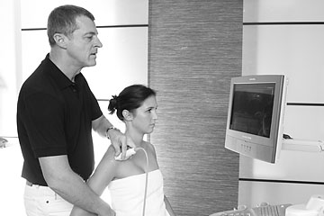
Ultrasound diagnostics
Sonography, also known as ultrasound diagnostics, is an established imaging technique for examining the musculoskeletal system. It enables the detailed visualisation of soft tissue structures such as muscles, tendons, ligaments and joints. The use of standardised examination techniques is essential for a precise diagnosis. Compared to other imaging methods, sonography has the advantage of not using radiation and of enabling dynamic examinations in real time. This makes it particularly valuable for assessing diseases and changes in muscles, tendons, soft tissues and joints.
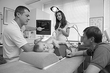
Early detection of hip dysplasia in infants
Ultrasound of the infant hip is an essential part of the U3 check-up, which is carried out in the 4th to 6th week of life. This examination enables the early detection of hip dysplasia, a malformation of the hip joint that can lead to serious health problems if left untreated.
Today, ultrasound monitoring of the infant hip is a must.
Limitations of sonography
- limited penetration depth: in obese patients or when organs are located deep inside, image quality can be reduced.
- Dependence on the examiner's experience: the quality and informative value of the examination depend to a large extent on the examiner's experience.
- Difficulties in visualising bony structures: ultrasound can only be used to a limited extent for assessing bones.
The advantages of sonography:
- No exposure: Neither the patient nor the examiner is exposed to ionising radiation, which makes the examination particularly gentle.
- Can be repeated as often as necessary: Since there is no radiation exposure, the examination can be repeated as often as necessary.
- Cost-effective: Compared to other imaging techniques, sonography is inexpensive and does not require a high level of technical expertise.
- Visualisation of motion sequences: real-time imaging allows the observation of dynamic processes, such as the movement of muscles and tendons.
- Side-by-side examination: Both sides of the body can be compared directly with each other to identify any differences.
- No metal artefacts: In contrast to computed tomography (CT) and magnetic resonance imaging (MRI), ultrasound does not produce any image interference caused by metallic implants.