

")
")
X-ray examination Berlin-Frohnau
An X-ray examination enables the visualisation of different body regions and tissues using X-rays. These rays were discovered in 1895 by the German physicist Wilhelm Conrad Röntgen.
How X-ray technology works
X-rays are high-energy electromagnetic waves that can penetrate matter. In medical diagnostics, they are used to visualise structures such as bones, organs and vessels. The rays are directed through the body and absorbed to varying degrees by the different tissues. This results in high-contrast images that help doctors make a diagnosis.
How an X-ray is taken
In an X-ray image, the body part to be examined is positioned between an X-ray source and a detector. The radiation source emits X-rays that penetrate the body. Denser tissue such as bone absorbs more radiation and appears lighter on the X-ray image, while softer tissue absorbs less radiation and appears darker. This principle enables the detailed visualisation of internal structures.
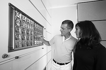
Evaluation of radiographs
The interpretation of radiographs requires a sound knowledge of anatomy and possible pathological changes. Particularly in children, it is important to consider the developmental stages of the skeleton in order to recognise growth disorders or deformities. Skeletal maturity is often determined by radiographs of the hand, whereby the ossification of the epiphyses is assessed.
Significance in orthopedics
In orthopedics, X-ray diagnostics play a central role, particularly in the planning of operations. Precise X-ray images can be used to accurately capture deformities and optimally prepare for surgical procedures. Modern digital planning procedures make it possible to select implants that fit perfectly and to plan the surgical steps in detail, which significantly influences the success of the treatment.
Radiation protection and modern developments
Despite the diagnostic advantages of X-rays, it is essential that we deal responsibly with radiation exposure. Modern equipment and techniques are designed to keep the radiation dose for patients as low as possible. In addition, advanced imaging techniques such as computed tomography (CT) or magnetic resonance imaging (MRI) complement the diagnostic process and, depending on the issue at hand, provide further detailed insights into the body.
We do not offer X-rays in Frohnau, but we recommend our specialist partner, the Röntgeninstitut Berlin at Möllendorffstr. 52, 10367 Berlin.